
Introduction — a quick scene, a few numbers, a pressing question
Have you ever stood over a lab bench and felt a small rush of doubt when an image didn’t match the story you expected? I have, and those moments teach us the most. laser speckle contrast imaging lsci can capture blood flow patterns across tissue in real time, often at tens to hundreds of frames per second and across thousands of pixels — that’s useful data, but it can be noisy. So how do we turn those shifting speckles into reliable, repeatable measures of perfusion that clinicians and researchers trust?
I’ll walk through what matters most: where the common methods trip up, where users quietly struggle, and which principles will help you get cleaner, faster, and more actionable results. Along the way I’ll be candid about trade-offs and practical fixes I use in the lab. (Yes, I’ve ruined plenty of datasets — and learned from each one.) Next, let’s dig into the hard part: why the usual solutions often fail us.

Deep Dive: Why Traditional Approaches Fall Short
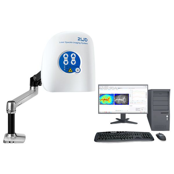
laser speckle contrast imaging system setups promise fast perfusion maps, but the reality is trickier. At its core, speckle contrast depends on dynamic scattering and exposure time. When exposure and frame rate don’t match the tissue dynamics, speckle contrast values skew. I say this from hands-on work: mismatched CMOS sensor settings and crude spatial filtering create artifacts that look like physiological change but are just measurement error.
What breaks first?
Two quick technical points explain a lot. First, frame rate vs. exposure trade-offs — push frame rate too high and you lose contrast; push exposure too long and motion blurs true flow. Second, sampling and sensor noise: low signal-to-noise on the sensor makes perfusion mapping unstable, especially near edges or in low-light tissue. These are not abstract issues. They cause false positives in studies, and they frustrate clinicians. Look, it’s simpler than you think: tune exposure and use modest spatial averaging rather than aggressive smoothing. Yes, really — small choices save entire experiments.
Forward View: Principles for Next-Gen LSCI
What should you build toward? Start with the measurement principles: match your exposure to the expected decorrelation time of the tissue, and favor sensors with stable gain and linear response. A modern laser speckle contrast imaging system pairs controlled illumination (laser diode stabilization) with a sensor pipeline that preserves phase information where possible. That combination reduces bias in speckle contrast and improves repeatability across sessions.
What’s Next — practical steps
First, automate a short calibration routine before each session. Second, integrate simple on-board preprocessing — dark-frame subtraction and basic flat-field correction — to cut sensor bias. Third, benchmark using a phantom with known flow rates, not just live tissue. These steps save time and make your perfusion maps meaningful. Also — funny how that works, right? — small investments in calibration yield the biggest gains in confidence.
To help you choose the right path, I suggest three evaluation metrics: 1) repeatability across repeated trials (look for low coefficient of variation), 2) sensitivity to known flow changes (can the system detect a 10% shift?), and 3) robustness to common artifacts (edge motion, ambient light changes). Use those to compare setups and claims. I’ve used these metrics when advising labs and they cut the noise quickly.
We want practical results, not neat figures on a slide. If you’re ready to move from noisy images to reliable perfusion mapping, these insights will guide you. For tools and validated systems, I often point teams toward proven vendors — and you can explore options at BPLabLine.